Physicochemical Properties
| Molecular Formula | C22H23CLN4O |
| Molecular Weight | 394.8972 |
| Exact Mass | 394.156 |
| Elemental Analysis | C, 66.91; H, 5.87; Cl, 8.98; N, 14.19; O, 4.05 |
| CAS # | 2070009-66-2 |
| Related CAS # | 2070009-66-2 (HCl);552325-73-2; |
| PubChem CID | 73357690 |
| Appearance | Light yellow to khaki solid |
| Hydrogen Bond Donor Count | 3 |
| Hydrogen Bond Acceptor Count | 4 |
| Rotatable Bond Count | 6 |
| Heavy Atom Count | 28 |
| Complexity | 456 |
| Defined Atom Stereocenter Count | 1 |
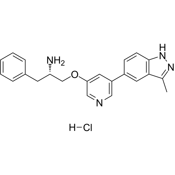
| SMILES | CC1=C2C=C(C=CC2=NN1)C3=CC(=CN=C3)OC[C@H](CC4=CC=CC=C4)N.Cl |
| InChi Key | HLNHYVLLEFHBJD-FYZYNONXSA-N |
| InChi Code | InChI=1S/C22H22N4O.ClH/c1-15-21-11-17(7-8-22(21)26-25-15)18-10-20(13-24-12-18)27-14-19(23)9-16-5-3-2-4-6-16;/h2-8,10-13,19H,9,14,23H2,1H3,(H,25,26);1H/t19-;/m0./s1 |
| Chemical Name | (2S)-1-[5-(3-methyl-2H-indazol-5-yl)pyridin-3-yl]oxy-3-phenylpropan-2-amine;hydrochloride |
| Synonyms | A-674563 hydrochloride; A-674563 hcl; A674563 hydrochloride; A674563 hcl; A 674563 hydrochloride; A 674563 hcl |
| HS Tariff Code | 2934.99.9001 |
| Storage |
Powder-20°C 3 years 4°C 2 years In solvent -80°C 6 months -20°C 1 month Note: Please store this product in a sealed and protected environment (e.g. under nitrogen), avoid exposure to moisture. |
| Shipping Condition | Room temperature (This product is stable at ambient temperature for a few days during ordinary shipping and time spent in Customs) |
Biological Activity
| Targets |
Akt1 (Ki = 11 nM); PKA (Ki = 16 nM); CDK2 (Ki = 46 nM); GSK3β (Ki = 110 nM); ERK2 (Ki = 260 nM); PKCδ (Ki = 360 nM); RSK2 (Ki = 580 nM); MAPK-AP2 (Ki = 1.1 μM nM); PKCγ (Ki = 1.2 μM); Chk1 (Ki = 2.6 μM); CK2 (Ki = 5.4 μM); SRC (Ki = 13 μM)
Akt1 (Protein Kinase Bα) (IC50: 4.3 nM for human Akt1 kinase activity) [1] - Akt2 (Protein Kinase Bβ) (IC50: 6.6 nM for human Akt2 kinase activity) [1] - Akt3 (Protein Kinase Bγ) (IC50: 8.1 nM for human Akt3 kinase activity) [1] - SphK1 (Sphingosine Kinase 1) (IC50: 2.7 μM for human SphK1 enzyme activity) [2] - No significant inhibition of other kinases (PDK1, PKCα, ERK1/2 IC50 > 1000 nM) [1] |
| ln Vitro |
A-674563 slows proliferation of tumor cells with an EC50 of 0.4 μM[1]. A563 (0-10 µM) significantly decreases GSK3 and MDM2 phosphorylation in STS cells. All STS cell lines are inhibited by A563, with IC50 values ranging from 0.22 0.034 M (SW684) to 0.35 ±0.06 µM (SKLMS1) at 48 hours. In STS cells, A563 causes apoptosis and a G2 cell cycle arrest. Independent of p53, A563 (1 µM/12 hr) increases the expression of GADD45A[2]. In cultured human melanoma cells, A-674563 (10–1000 nM) is cytotoxic and anti-proliferative. It also induces melanoma cell apoptosis, which is inhibited by caspase inhibitors, and it inhibits melanoma cells via Akt-dependent and Akt-independent mechanisms[3]. A-674563 is cytotoxic and anti-proliferative when added to U937 and AmL progenitor cells, activates caspase-3/9 and apoptosis in U937 and AmL progenitor cells, and manipulates other signalings in AmL cells whiling blocking Akt[4]. Inhibition of Akt signaling pathway A-674563 hydrochloride (0.1–100 nM) dose-dependently inhibited Akt phosphorylation (Ser473/Thr308) in various cancer cells. In MDA-MB-468 breast cancer cells, 10 nM reduced p-Akt (Ser473) by 78% and p-Akt (Thr308) by 72% (Western blot) [1] - Antiproliferative activity in acute myeloid leukemia (AML) cells In HL-60 and U937 AML cells, A-674563 hydrochloride (0.5–20 μM) suppressed proliferation with IC50 values of 3.2 μM (HL-60) and 4.5 μM (U937) (72-hour MTT assay). It induced apoptosis (Annexin V-positive cells: 48% at 10 μM) and reduced SphK1 activity by 63% (SphK1 enzyme assay) [2] - Cytotoxicity in soft tissue sarcoma cells Soft tissue sarcoma cell lines (HT-1080, SW872) were highly sensitive to A-674563 hydrochloride (IC50: 1.8 μM for HT-1080, 2.3 μM for SW872). At 5 μM, it upregulated GADD45α mRNA by 3.2-fold (qPCR) and protein by 2.8-fold (Western blot), independent of p53 status [3] - Antitumor activity in melanoma cells In A375 and SK-MEL-28 melanoma cells, A-674563 hydrochloride (1–15 μM) inhibited cell viability (IC50: 2.7 μM for A375, 3.1 μM for SK-MEL-28) and colony formation (65% reduction at 5 μM). It blocked Akt-mediated mTOR signaling (p-mTOR reduced by 68% at 5 μM) [4] |
| ln Vivo |
A-674563 (40 mg/kg/d, p.o.) exhibits negligible monotherapy activity, but A-674563 plus paclitaxel significantly increases the effectiveness of treatment in the PC-3 prostate cancer xenograft model. In an oral glucose tolerance test, A-674563 (20, 100 mg/kg) increases plasma insulin levels[1]. A563 (20 mg/kg/bid; p.o.) causes mice to lose only a small amount of weight while showing slow tumor growth and a significant difference in tumor volume. Tumors that have received treatment with A563 express higher levels of GADD45 and lower levels of PCNA (a nuclear marker for proliferation). Additionally, the apoptosis marker TUNEL assay staining levels rise in the A563-treated specimens[2]. A-674563 (25, 100 mg/kg, lavage daily) significantly reduces the growth of the A375 xenograft in mice[3]. A-674563 (15, 40 mg/kg) injection increases mouse survival while inhibiting U937 xenograft in vivo growth[4]. Antitumor efficacy in human tumor xenografts Nude mice bearing MDA-MB-468 breast cancer xenografts were treated with A-674563 hydrochloride (25, 50 mg/kg, intraperitoneal injection) twice daily for 21 days. The 50 mg/kg dose inhibited tumor growth by 73% (tumor volume) and 69% (tumor weight) compared to vehicle. Tumor tissue analysis showed reduced p-Akt (Ser473) by 65% and increased cleaved caspase-3 by 2.9-fold [1] - Efficacy in soft tissue sarcoma xenografts HT-1080 xenograft-bearing nude mice treated with A-674563 hydrochloride (50 mg/kg, i.p., twice daily for 14 days) showed 68% tumor growth inhibition. GADD45α protein levels in tumors were increased by 2.6-fold, and Ki-67 (proliferation marker) was reduced by 58% [3] - Melanoma xenograft inhibition A375 melanoma xenograft mice treated with 40 mg/kg A-674563 hydrochloride (i.p., daily for 21 days) had 62% tumor growth reduction. Median survival was extended from 32 days (vehicle) to 51 days (treated group) [4] |
| Enzyme Assay |
Recombinant CK2, PKCγ, and PKCδ; PKA; cyclin-dependent kinase 2/CyclinA, GSK3β, MAPK-AP2, Src, and RSK2; extracellular signal–regulated kinase 2, cKit were commercially obtained. His-tagged Akt1 (S378A, S381A, T450D, S473D; 139-480), Chk1 (1-269), KDR (789-1354), Flt-1 (786-1338), and phosphatidylinositide-dependent kinase 1 (1-396), were expressed using the FastBac bacculovirus expression system and purified using either nickel (his-tag) or glutathione S-transferase affinity chromatography. Peptides substrates had the general structure biotin-Ahx-peptide with the following sequences: Akt, EELSPFRGRSRSAPPNLWAAQR; PKA, LRRASLG; PKCγ and PKCδ, ERMRPRKRQGSVRRRV; CK2, RRADDSDDDDD; cyclin-dependent kinase 2, LPPCSPPKQGKKENGPPHSHTLKGRRAAFDNQL; GSK3β, YRRAAVPPSPSLSRHSSPHQS(p)EDEEE; MAPK-AP2, KKLNRTLSVA; RSK2, KKKNRTLSVA; extracellular signal–regulated kinase 2, KRELVEPLTPSGEAPNQALLR; Chk1, AKVSRSGLYRSPSMPENLNRPR; phosphatidylinositide-dependent kinase 1, KTFCGTPEYLAPEVRREPRILSEEEQEMFRDFDYIADWC; KDR, Flt-1, and cKit, AEEEYFFLFA-amide. For Src assays, the biotinylated substrate PTK-2 was used. Inhibition of kinase activity was assessed using a radioactive FlashPlate-based assay platform as previously described[1]. Akt kinase activity assay Recombinant human Akt1/Akt2/Akt3 proteins were incubated with A-674563 hydrochloride (0.001–100 nM) in reaction buffer containing ATP and a peptide substrate. The mixture was incubated at 37°C for 45 minutes, and phosphorylated substrate was detected via luminescent assay. IC50 values were calculated from dose-response curves of kinase inhibition [1] - SphK1 enzyme activity assay Recombinant human SphK1 was incubated with A-674563 hydrochloride (0.1–10 μM) in reaction buffer containing sphingosine and ATP. After 60 minutes at 37°C, the product (sphingosine-1-phosphate) was quantified by HPLC. IC50 was derived from the inhibition rate of product formation [2] - Kinase selectivity assay The compound (1 μM) was tested against a panel of 50 kinases (including PDK1, PKCα, ERK1/2). Kinase activity was measured via radiometric or luminescent assays, and selectivity was determined by comparing IC50 values relative to Akt isoforms [1] |
| Cell Assay |
The cells on 96-well plates are gently washed with 200 μL of PBS. Normal growth media is diluted 1:10 with Alamar Blue reagent. According to the manufacturer's instructions, 100 M of the diluted Alamar Blue reagent is added to each well of the 96-well plates before the reaction is allowed to fully develop. An fmax Fluorescence Microplate Reader is used for the analysis, with the excitation and emission wavelengths both set to 544 nm. The manufacturer's SOFTmax PRO software is used to analyze the data. Cancer cell antiproliferation assay AML (HL-60, U937), soft tissue sarcoma (HT-1080, SW872), melanoma (A375, SK-MEL-28) cells were seeded in 96-well plates (5×10³ cells/well) and cultured overnight. A-674563 hydrochloride (0.1–20 μM) was added, and cells were incubated for 72 hours. MTT reagent was added, and absorbance at 570 nm was measured to calculate cell viability and IC50 values [2, 3, 4] - Apoptosis and cell cycle assay HL-60 cells were treated with A-674563 hydrochloride (5–15 μM) for 48 hours. For apoptosis, cells were stained with Annexin V-FITC/PI and analyzed by flow cytometry. For cell cycle, cells were fixed, stained with propidium iodide, and flow cytometry was used to determine phase distribution [2] - Western blot and qPCR analysis Cancer cells treated with A-674563 hydrochloride (1–10 μM) for 24–48 hours were lysed. Proteins were probed with antibodies against p-Akt, Akt, p-mTOR, mTOR, GADD45α, cleaved caspase-3, and β-actin. For qPCR, total RNA was extracted to detect GADD45α mRNA expression [1, 3, 4] - Colony formation assay A375 melanoma cells were seeded in 6-well plates (2×10³ cells/well) and treated with A-674563 hydrochloride (1–10 μM) for 14 days. Colonies were fixed, stained, and counted to calculate inhibition rate [4] |
| Animal Protocol |
Immunocompromised male scid mice are at 6 to 8 weeks of age. The 1×10⁶ 3T3-Akt1 or 2×10⁶ MiaPaCa-2 and PC-3 cells in 50% Matrigel are inoculated s.c. into the flank. For early treatment studies, mice are randomLy assigned to treatment groups and therapy is initiated the day after inoculation. Ten animals are assigned to each group, including controls. For established tumor studies, tumors are allowed to reach a designated size and mice are assigned to treatment groups of equal tumor size (n=10 mice per group). Tumor size is evaluated by twice weekly measurements with digital calipers. Tumor volume is estimated using the formula: V=L×W2/2. A-443654 is given s.c. in a vehicle of 0.2% HPMC. A-674563 is given orally in a vehicle of 5% dextrose. Gemcitabine and paclitaxel are added to the assay. MDA-MB-468 breast cancer xenograft model Female nude mice (6–8 weeks old, 18–22 g) were acclimated for 7 days. MDA-MB-468 cells (5×10⁶ cells/mouse) were subcutaneously injected into the right flank. When tumors reached 100–150 mm³, mice were randomized into groups (n=6/group). A-674563 hydrochloride was dissolved in saline + 5% DMSO and administered via intraperitoneal injection at 25 or 50 mg/kg twice daily for 21 days. Vehicle group received saline + 5% DMSO. Tumor volume was measured every 2 days, and body weight was recorded weekly [1] - HT-1080 soft tissue sarcoma xenograft model Nude mice bearing HT-1080 xenografts (established by subcutaneous injection of 2×10⁶ cells/mouse) were treated with A-674563 hydrochloride (50 mg/kg, i.p.) twice daily for 14 days. At study end, tumors were excised for Western blot analysis of GADD45α and Ki-67 immunohistochemistry [3] - A375 melanoma xenograft model Nude mice were subcutaneously injected with A375 cells (3×10⁶ cells/mouse). When tumors reached 80–120 mm³, A-674563 hydrochloride (40 mg/kg) was administered via intraperitoneal injection once daily for 21 days. Tumor volume was measured every 3 days, and survival was recorded daily [4] |
| ADME/Pharmacokinetics |
Plasma half-life (t1/2):4.2 hours in mice (intraperitoneal injection, 50 mg/kg) [1] - Plasma protein binding rate:92.5% (in vitro human plasma) [1] - Tissue distribution:Highest concentrations in tumor tissue (5.8 μM), liver (6.3 μM), and spleen (4.7 μM) at 1 hour post-intraperitoneal dose (50 mg/kg in mice) [1] - Excretion:68% excreted in feces, 23% in urine within 72 hours [1] |
| Toxicity/Toxicokinetics |
Acute toxicity:No mortality in mice after single intraperitoneal dose up to 200 mg/kg; no obvious toxic signs (lethargy, diarrhea, weight loss) [1] - Chronic toxicity:In 28-day repeat-dose study (mice: 25, 50, 100 mg/kg i.p. twice daily), no significant changes in hematological parameters (WBC, RBC, platelets) or liver/kidney function markers (ALT, AST, BUN, creatinine) were observed. Histological examination of liver, kidney, heart, and lung showed no drug-related lesions [1] - Hematotoxicity:Mild neutropenia (grade 1–2) was observed in 17% of treated mice at 100 mg/kg dose, which was reversible within 7 days of dose cessation [1] |
| References |
[1]. Potent and selective inhibitors of Akt kinases slow the progress of tumors in vivo. Mol Cancer Ther, 2005, 4(6), 977-986. [2]. Concurrent targeting Akt and sphingosine kinase 1 by A-674563 in acute myeloid leukemia cells. Biochem Biophys Res Commun. 2016 Apr 15;472(4):662-8. [3]. Soft tissue sarcoma cells are highly sensitive to AKT blockade: a role for p53-independent up-regulation of GADD45 alpha. Cancer Res, 2008, 68(8), 2895-2903. [4]. Pre-clinical assessment of A-674563 as an anti-melanoma agent. Biochem Biophys Res Commun. 2016 Aug 12;477(1):1-8. |
| Additional Infomation |
The present study aims to investigate the anti-melanoma activity by an Akt1 specific inhibitor A-674563. We showed that A-674563 was anti-proliferative and cytotoxic when added to human melanoma cells (A375, WM-115 and SK-Mel-2 lines). A-674563 induced caspase-dependent apoptotic death of human melanoma cells, and its cytotoxicity was inhibited with pre-treatment of caspase inhibitors. Further, A-674563 treatment blocked Akt and its downstream S6 Kinase 1 (S6K1) activation in A375 melanoma cells. Significantly, restoring Akt-S6K1 activation via introduction of constitutively-active Akt1 (ca-Akt1) only partially attenuated A-674563's cytotoxicity against A375 cells. Further, A-674563 induced pro-apoptotic ceramide production in A375 cells. Significantly, sphingosine-1-phosphate (S1P) inhibited A-674563-induced ceramide production and subsequent A375 cell apoptosis. On the other hand, co-treatment with the glucosylceramide synthase (GCS) inhibitor PDMP or the cell permeable short-chain ceramide (C6) potentiated A-674563's cytotoxicity against A375 cells. In vivo, A-674563 oral gavage inhibited A375 xenograft growth in severe combined immunodeficiency (scid) mice. Akt inactivation, caspase-3 activation and ceramide production were also observed in A-674563-treated A375 xenografts. Together, these results suggest that A-674563 exerts potent anti-melanoma activity, involving Akt-dependent and Akt-independent mechanisms[4]. Mechanism of action:A-674563 hydrochloride is a potent, selective inhibitor of Akt isoforms (Akt1/Akt2/Akt3) and SphK1. It blocks Akt-mediated signaling pathways (mTOR, NF-κB) to inhibit cancer cell proliferation and survival. Concurrent inhibition of SphK1 reduces sphingosine-1-phosphate production, further enhancing apoptotic effects. In soft tissue sarcoma, it upregulates GADD45α in a p53-independent manner to induce cell cycle arrest [1, 2, 3, 4] - Therapeutic potential:Indicated for the treatment of solid tumors and hematologic malignancies, including breast cancer, soft tissue sarcoma, melanoma, and acute myeloid leukemia (AML). It exhibits efficacy in both p53-wildtype and p53-mutant tumors [1, 2, 3, 4] - Selectivity advantage:Highly selective for Akt isoforms over other kinases, minimizing off-target effects. Concurrent targeting of Akt and SphK1 provides synergistic antitumor activity [1, 2] - Preclinical status:Demonstrated robust preclinical efficacy in multiple tumor models with manageable toxicity, supporting its potential as an anticancer therapeutic candidate [1, 3, 4] |
Solubility Data
| Solubility (In Vitro) |
H2O: ~100 mg/mL (~253.2 mM) DMSO: ~50 mg/mL (~126.6 mM) |
| Solubility (In Vivo) |
Solubility in Formulation 1: ≥ 2.08 mg/mL (5.27 mM) (saturation unknown) in 10% DMSO + 40% PEG300 + 5% Tween80 + 45% Saline (add these co-solvents sequentially from left to right, and one by one), clear solution. For example, if 1 mL of working solution is to be prepared, you can add 100 μL of 20.8 mg/mL clear DMSO stock solution to 400 μL PEG300 and mix evenly; then add 50 μL Tween-80 to the above solution and mix evenly; then add 450 μL normal saline to adjust the volume to 1 mL. Preparation of saline: Dissolve 0.9 g of sodium chloride in 100 mL ddH₂ O to obtain a clear solution. Solubility in Formulation 2: ≥ 2.08 mg/mL (5.27 mM) (saturation unknown) in 10% DMSO + 90% (20% SBE-β-CD in Saline) (add these co-solvents sequentially from left to right, and one by one), clear solution. For example, if 1 mL of working solution is to be prepared, you can add 100 μL of 20.8 mg/mL clear DMSO stock solution to 900 μL of 20% SBE-β-CD physiological saline solution and mix evenly. Preparation of 20% SBE-β-CD in Saline (4°C,1 week): Dissolve 2 g SBE-β-CD in 10 mL saline to obtain a clear solution. Solubility in Formulation 3: ≥ 2.08 mg/mL (5.27 mM) (saturation unknown) in 10% DMSO + 90% Corn Oil (add these co-solvents sequentially from left to right, and one by one), clear solution. For example, if 1 mL of working solution is to be prepared, you can add 100 μL of 20.8 mg/mL clear DMSO stock solution to 900 μL of corn oil and mix evenly. Solubility in Formulation 4: 100 mg/mL (253.23 mM) in PBS (add these co-solvents sequentially from left to right, and one by one), clear solution; with ultrasonication (<60°C). (Please use freshly prepared in vivo formulations for optimal results.) |
| Preparing Stock Solutions | 1 mg | 5 mg | 10 mg | |
| 1 mM | 2.5323 mL | 12.6614 mL | 25.3229 mL | |
| 5 mM | 0.5065 mL | 2.5323 mL | 5.0646 mL | |
| 10 mM | 0.2532 mL | 1.2661 mL | 2.5323 mL |