Physicochemical Properties
| Molecular Formula | C5H5N5O2 |
| Molecular Weight | 167.1255 |
| Exact Mass | 167.044 |
| CAS # | 5614-64-2 |
| PubChem CID | 135420630 |
| Appearance | Light brown to khaki solid powder |
| Density | 2.49g/cm3 |
| Boiling Point | 327.7ºC at 760 mmHg |
| Melting Point | >400ºC dec. |
| Flash Point | 152ºC |
| Index of Refraction | 2.156 |
| LogP | -2.2 |
| Hydrogen Bond Donor Count | 4 |
| Hydrogen Bond Acceptor Count | 3 |
| Rotatable Bond Count | 0 |
| Heavy Atom Count | 12 |
| Complexity | 342 |
| Defined Atom Stereocenter Count | 0 |
| InChi Key | CLGFIVUFZRGQRP-UHFFFAOYSA-N |
| InChi Code | InChI=1S/C5H5N5O2/c6-4-8-2-1(3(11)10-4)7-5(12)9-2/h(H5,6,7,8,9,10,11,12) |
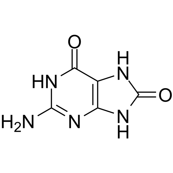
| Chemical Name | 2-amino-7,9-dihydro-1H-purine-6,8-dione |
| HS Tariff Code | 2934.99.9001 |
| Storage |
Powder-20°C 3 years 4°C 2 years In solvent -80°C 6 months -20°C 1 month |
| Shipping Condition | Room temperature (This product is stable at ambient temperature for a few days during ordinary shipping and time spent in Customs) |
Biological Activity
| Targets |
- DNA (induces G→T and A→C base substitutions by disrupting normal DNA base pairing; no IC50/Ki/EC50 data available) [2], [3] - 8-Oxoguanine DNA Glycosylase (OGG1) (serves as a substrate for OGG1-mediated DNA repair; no IC50/Ki/EC50 data available) [1], [2] |
| ln Vitro |
- In oligonucleotide-based replication assays, 8-Hydroxyguanine caused significant base mispairing during DNA synthesis. When incorporated into a 20-mer oligonucleotide (position 10 as 8-hydroxyguanine) and used as a template for DNA polymerase α, it induced G→T substitutions at a frequency of 32% (vs. <0.1% for unmodified guanine) and A→C substitutions at 18% (measured by Sanger sequencing of replication products) [3] - In human embryonic kidney (HEK293T) cells transfected with plasmids containing 8-Hydroxyguanine (within the neomycin resistance gene), the mutation frequency of the neomycin gene increased by 8.5-fold compared to plasmids with unmodified guanine (12.6×10⁻⁴ vs. 1.5×10⁻⁴ mutations/base, determined by selecting G418-resistant colonies and sequencing) [3] - In vitro OGG1 repair assays showed that 8-Hydroxyguanine was recognized and excised by recombinant OGG1. Incubation of 8-hydroxyguanine-containing DNA (50 ng) with OGG1 (10 U) for 30 min at 37°C resulted in 75% cleavage of the damaged DNA (detected by 12% polyacrylamide gel electrophoresis and ethidium bromide staining) [1] |
| ln Vivo |
- In Mmh/Ogg1-deficient mice (OGG1 knockout, unable to repair 8-Hydroxyguanine), chronic oxidative stress (induced by intraperitoneal injection of D-galactosamine, 200 mg/kg twice weekly for 12 weeks) led to a 4.2-fold increase in 8-Hydroxyguanine levels in liver DNA (18.6 vs. 4.4 lesions/10⁶ guanines in wild-type mice) and a 3.8-fold increase in kidney DNA (16.2 vs. 4.2 lesions/10⁶ guanines) [1] - In wild-type C57BL/6 mice exposed to chronic cigarette smoke (6 cigarettes/day, 5 days/week for 6 months), 8-Hydroxyguanine accumulated in lung DNA (9.8 lesions/10⁶ guanines vs. 3.1 in control mice) and was associated with a 2.5-fold higher incidence of lung adenomas at 18 months (p < 0.05) [2] - In Ogg1-deficient mice without external oxidative stress, basal 8-Hydroxyguanine levels in brain DNA were 2.1-fold higher than in wild-type mice (7.3 vs. 3.5 lesions/10⁶ guanines), and these mice showed a 30% higher spontaneous liver tumor rate at 24 months [1] |
| Enzyme Assay |
- OGG1 activity assay for 8-Hydroxyguanine repair: 1) Prepare double-stranded DNA substrate (40 bp) containing a single 8-Hydroxyguanine residue at position 20 (paired with cytosine). 2) Incubate 50 ng of the substrate with recombinant OGG1 (0-20 U) in reaction buffer (50 mM Tris-HCl pH 7.5, 10 mM EDTA, 1 mM DTT) at 37°C for 30 min. 3) Stop the reaction by adding 0.5% SDS and 10 mM EDTA, then treat with proteinase K (50 μg/mL) at 55°C for 15 min. 4) Separate the DNA products by 12% polyacrylamide gel electrophoresis, stain with ethidium bromide, and quantify the cleaved (single-stranded) DNA band intensity to calculate OGG1 activity [1] - DNA polymerase misincorporation assay: 1) Synthesize a 30-mer template oligonucleotide with 8-Hydroxyguanine at position 15. 2) Incubate the template (1 μM) with DNA polymerase α (5 U), dNTPs (100 μM each), and reaction buffer (20 mM Tris-HCl pH 7.9, 10 mM MgCl₂, 50 mM KCl) at 37°C for 60 min. 3) Purify the newly synthesized DNA strands by ethanol precipitation, then sequence them using Sanger sequencing. 4) Count the number of G→T and A→C substitutions at the 8-Hydroxyguanine site to calculate misincorporation frequency [3] |
| Cell Assay |
- 8-Hydroxyguanine mutation frequency assay in mammalian cells: 1) Construct a pSV2neo plasmid containing a single 8-Hydroxyguanine residue within the neomycin resistance gene (position 456). 2) Transfect the plasmid (2 μg) into CHO-K1 cells using lipofection, and select G418-resistant colonies (600 μg/mL G418) for 14 days. 3) Pick 50 resistant colonies per group, extract plasmid DNA from each colony, and sequence the neomycin resistance gene. 4) Calculate the mutation frequency as (number of colonies with G→T/A→C substitutions) / total number of sequenced colonies [3] - Intracellular 8-Hydroxyguanine detection assay: 1) Treat HEK293T cells with H₂O₂ (100 μM) for 2 hours to induce oxidative DNA damage. 2) Harvest cells, extract genomic DNA using phenol-chloroform method, and digest 10 μg of DNA with DNase I and alkaline phosphatase. 3) Separate the nucleosides by high-performance liquid chromatography (HPLC) with electrochemical detection (ECD, 0.6 V potential). 4) Quantify 8-Hydroxyguanine levels by comparing the peak area to a standard curve (0.1-10 ng/mL 8-Hydroxyguanine standard) [1] |
| Animal Protocol |
- Ogg1-deficient mouse chronic oxidative stress experiment: 1) Use Mmh/Ogg1⁻/⁻ mice (C57BL/6 background, 8-week-old, n=10 per group) and wild-type (Ogg1⁺/⁺) littermates as controls. 2) Induce chronic oxidative stress by intraperitoneal injection of D-galactosamine (200 mg/kg) twice weekly for 12 weeks; control groups receive saline injections. 3) At the end of treatment, euthanize mice, collect liver, kidney, and brain tissues. 4) Extract genomic DNA from each tissue (100 mg), digest with enzymes, and quantify 8-Hydroxyguanine levels by HPLC-ECD. 5) For long-term tumor monitoring, keep a subset of mice (n=15 per group) for 24 months, perform monthly palpation to detect liver tumors, and confirm with histopathology at euthanasia [1] - Wild-type mouse cigarette smoke exposure experiment: 1) Use 6-week-old C57BL/6 mice (n=20 per group). 2) Expose mice to mainstream cigarette smoke (6 cigarettes/day, 5 days/week) in a whole-body exposure chamber for 6 months; control mice are exposed to filtered air. 3) At 6 months, collect lung tissue to measure 8-Hydroxyguanine levels via HPLC-ECD. 4) Continue housing mice for 12 more months (total 18 months), sacrifice, and examine lung tissues for adenomas via hematoxylin-eosin staining [2] |
| Toxicity/Toxicokinetics |
- Mutagenicity: 8-Hydroxyguanine is a potent mutagen. In vitro, it induces G→T and A→C substitutions with a frequency of 12-32% (depending on DNA polymerase type) [3]. In vivo, its accumulation in Ogg1-deficient mice increases spontaneous mutation rates by 2.1-4.2-fold in various tissues (liver, kidney, brain) [1] - Carcinogenicity: Chronic accumulation of 8-Hydroxyguanine is associated with increased cancer risk. Ogg1-deficient mice show a 30% higher spontaneous liver tumor rate at 24 months, and wild-type mice exposed to cigarette smoke (with elevated lung 8-Hydroxyguanine) have a 2.5-fold higher lung adenoma incidence at 18 months [1], [2] - No traditional "drug toxicity" data (e.g., LD50, hepatotoxicity) available, as 8-Hydroxyguanine is an endogenous oxidative DNA damage product, not a therapeutic drug [1], [2], [3] |
| References |
[1]. High accumulation of oxidative DNA damage, 8-hydroxyguanine, in Mmh/Ogg1 deficient mice by chronic oxidative stress. Carcinogenesis. 2002 Dec;23(12):2005-10. [2]. The role of 8-hydroxyguanine in carcinogenesis. Carcinogenesis. 1990 Sep;11(9):1447-50. [3]. 8-Hydroxyguanine, an abundant form of oxidative DNA damage, causes G----T and A----C substitutions. J Biol Chem. 1992 Jan 5;267(1):166-72. |
| Additional Infomation |
7,8-dihydro-8-oxoguanine is an oxopurine that is guanine in which the hydrogen at position 8 is replaced by an oxo group and in which the nitrogens at positions 7 and 9 each bear a hydrogen. It is functionally related to a guanine. 8-Hydroxyguanine has been reported in Caenorhabditis elegans with data available. - 8-Hydroxyguanine is the most abundant and well-characterized oxidative DNA damage product, formed endogenously by reactive oxygen species (ROS) or exogenously by environmental stressors (e.g., cigarette smoke, radiation) [1], [2] - Its carcinogenic mechanism involves escaping DNA repair (e.g., OGG1 deficiency) and causing base mispairing during DNA replication: 8-Hydroxyguanine preferentially pairs with adenine instead of cytosine, leading to G→T transversions, and can also induce A→C substitutions in complementary strands [2], [3] - It is widely used as a biomarker for oxidative stress and DNA damage in studies of cancer, aging, and neurodegenerative diseases. Higher 8-Hydroxyguanine levels in tissues correlate with increased disease risk [1], [2] - 8-Hydroxyguanine is not a therapeutic drug; its study focuses on understanding oxidative DNA damage-related pathogenesis and developing repair-targeted therapies (e.g., OGG1 activators) [1], [2] |
Solubility Data
| Solubility (In Vitro) |
1M NaOH : 25 mg/mL (~149.58 mM) H2O : ~4.72 mg/mL (~28.24 mM) Ethanol :< 1 mg/mL DMSO :< 1 mg/mL |
| Solubility (In Vivo) |
Note: Listed below are some common formulations that may be used to formulate products with low water solubility (e.g. < 1 mg/mL), you may test these formulations using a minute amount of products to avoid loss of samples. Injection Formulations (e.g. IP/IV/IM/SC) Injection Formulation 1: DMSO : Tween 80: Saline = 10 : 5 : 85 (i.e. 100 μL DMSO stock solution → 50 μL Tween 80 → 850 μL Saline) *Preparation of saline: Dissolve 0.9 g of sodium chloride in 100 mL ddH ₂ O to obtain a clear solution. Injection Formulation 2: DMSO : PEG300 :Tween 80 : Saline = 10 : 40 : 5 : 45 (i.e. 100 μL DMSO → 400 μLPEG300 → 50 μL Tween 80 → 450 μL Saline) Injection Formulation 3: DMSO : Corn oil = 10 : 90 (i.e. 100 μL DMSO → 900 μL Corn oil) Example: Take the Injection Formulation 3 (DMSO : Corn oil = 10 : 90) as an example, if 1 mL of 2.5 mg/mL working solution is to be prepared, you can take 100 μL 25 mg/mL DMSO stock solution and add to 900 μL corn oil, mix well to obtain a clear or suspension solution (2.5 mg/mL, ready for use in animals). Injection Formulation 4: DMSO : 20% SBE-β-CD in saline = 10 : 90 [i.e. 100 μL DMSO → 900 μL (20% SBE-β-CD in saline)] *Preparation of 20% SBE-β-CD in Saline (4°C,1 week): Dissolve 2 g SBE-β-CD in 10 mL saline to obtain a clear solution. Injection Formulation 5: 2-Hydroxypropyl-β-cyclodextrin : Saline = 50 : 50 (i.e. 500 μL 2-Hydroxypropyl-β-cyclodextrin → 500 μL Saline) Injection Formulation 6: DMSO : PEG300 : castor oil : Saline = 5 : 10 : 20 : 65 (i.e. 50 μL DMSO → 100 μLPEG300 → 200 μL castor oil → 650 μL Saline) Injection Formulation 7: Ethanol : Cremophor : Saline = 10: 10 : 80 (i.e. 100 μL Ethanol → 100 μL Cremophor → 800 μL Saline) Injection Formulation 8: Dissolve in Cremophor/Ethanol (50 : 50), then diluted by Saline Injection Formulation 9: EtOH : Corn oil = 10 : 90 (i.e. 100 μL EtOH → 900 μL Corn oil) Injection Formulation 10: EtOH : PEG300:Tween 80 : Saline = 10 : 40 : 5 : 45 (i.e. 100 μL EtOH → 400 μLPEG300 → 50 μL Tween 80 → 450 μL Saline) Oral Formulations Oral Formulation 1: Suspend in 0.5% CMC Na (carboxymethylcellulose sodium) Oral Formulation 2: Suspend in 0.5% Carboxymethyl cellulose Example: Take the Oral Formulation 1 (Suspend in 0.5% CMC Na) as an example, if 100 mL of 2.5 mg/mL working solution is to be prepared, you can first prepare 0.5% CMC Na solution by measuring 0.5 g CMC Na and dissolve it in 100 mL ddH2O to obtain a clear solution; then add 250 mg of the product to 100 mL 0.5% CMC Na solution, to make the suspension solution (2.5 mg/mL, ready for use in animals). Oral Formulation 3: Dissolved in PEG400 Oral Formulation 4: Suspend in 0.2% Carboxymethyl cellulose Oral Formulation 5: Dissolve in 0.25% Tween 80 and 0.5% Carboxymethyl cellulose Oral Formulation 6: Mixing with food powders Note: Please be aware that the above formulations are for reference only. InvivoChem strongly recommends customers to read literature methods/protocols carefully before determining which formulation you should use for in vivo studies, as different compounds have different solubility properties and have to be formulated differently. (Please use freshly prepared in vivo formulations for optimal results.) |
| Preparing Stock Solutions | 1 mg | 5 mg | 10 mg | |
| 1 mM | 5.9834 mL | 29.9168 mL | 59.8337 mL | |
| 5 mM | 1.1967 mL | 5.9834 mL | 11.9667 mL | |
| 10 mM | 0.5983 mL | 2.9917 mL | 5.9834 mL |